 Fraunhofer Institute for Digital Medicine MEVIS
Aimed at the inspection of changes in cardiac function due to influences such as breathing or arrhythmia, the software suite CaFuR provides advanced segmentation, registration and exploration techniques. These features enable the automatic calculation of functional cardiac parameters per heart cycle as well as their variation over time. Automated analysis, modularity, data scalability and the advanced multi-cycle data exploration set CaFuR apart as a cutting-edge software solution for the efficient analysis of real-time cardiac-MRI.
Multicycle Real-time MRI Analysis
CaFuR enables cardiac diagnosis with real-time MRI. It consists of an accelerated MRI acquisition and a complete software solution for automated analysis.
© Fraunhofer MEVIS
Cardiac Function in Real-time (CaFuR)
Real-time MRI Analysis - Automatic, Quantitative and Multi-cycle
Quantitative Assessment of Functional Variability with Real-time MRI

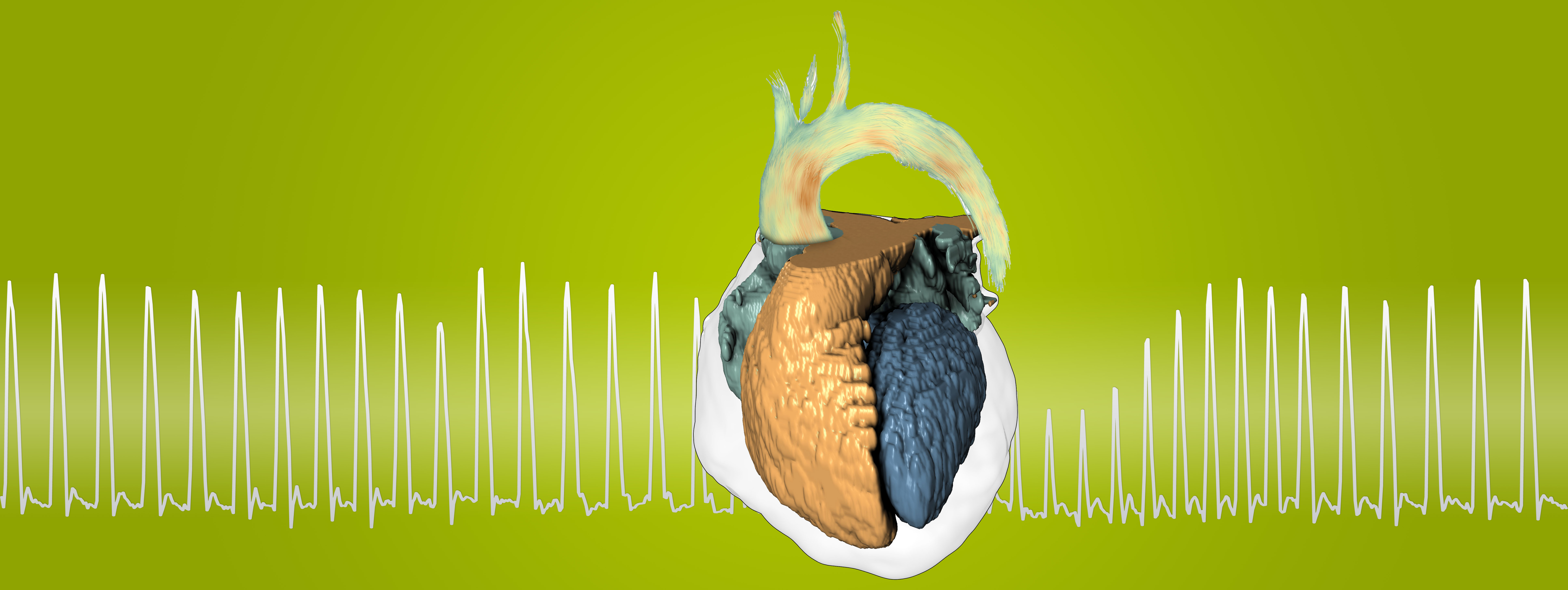
Schematic structure of the heart. The time diagram in the background shows the blood volume in the left ventricle (left) and the blood flow in the aortic arch (right).
Magnetic Resonance Imaging (MRI) is the best diagnostic procedure for many cardiac conditions, but is complicated and expensive. Our software add-on to existing MRI scanners allows diagnosis on novel real-time imaging sequences, which simplifies the procedure, significantly reduces the cost per patient and provides additional diagnostic information.
Examination speed-up enabled by CaFuR
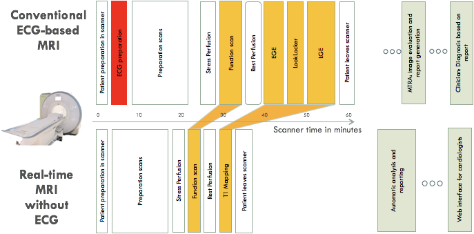
Conventional ECG-based MRI takes 60 or more minutes of scanner time and consists of a single heart-cycle analysis. CaFuR's real-time and ECG-less MRI is 40% faster and facilitate multi-cycle analysis. The automatic analysis takes just 2 min - compared to an interactive hours-long image analysis of a technician a huge advance.
Comprehensive Interface guides through the Workflow
- Automatic Myocardium and Vessel Segmentation & Tracking
- Automatic Cardiac Cycle Detection
- Quantitative Evaluation of Cardiac Flow & Function (End-Diastolic Fraction, End-Systolic Fraction, Stroke Volume, Ejection Fraction): Globally, Slicewise and Cyclewise
- Motion Analysis (by Elastic Registration): Radial Strain and Torsion – Wall Thickness
Publications
- Quantitative Assessment of Functional Variability with Real-time MRI
- Carotid artery flow as determined by real-time phase-contrast flow MRI and neurovascular ultrasound: A comparative study of healthy subjects
- Respiration and the watershed of spinal CSF flow in humans
- Exploration of Interventricular Septum Motion in Multi-Cycle Cardiac MRI
- Real-time myocardium segmentation for the assessment of cardiac function variation
- Identification of Upward movement of human CSF in Vivo and its relation to the Brain venous system
- Real-time magnetic resonance imaging of deep venous flow during muscular exercise—preliminary experience
- Advances in real‐time phase‐contrast flow MRI using asymmetric radial gradient echoes
- Automatic multi-cycle analysis of cardiac function from real-time MRI
- Analysis of aortic blood-flow from ECG-free realtime PC MRI
- Real-time magnetic resonance imaging of cardiac function and flow—recent progress