Fraunhofer Institute for Digital Medicine MEVIS
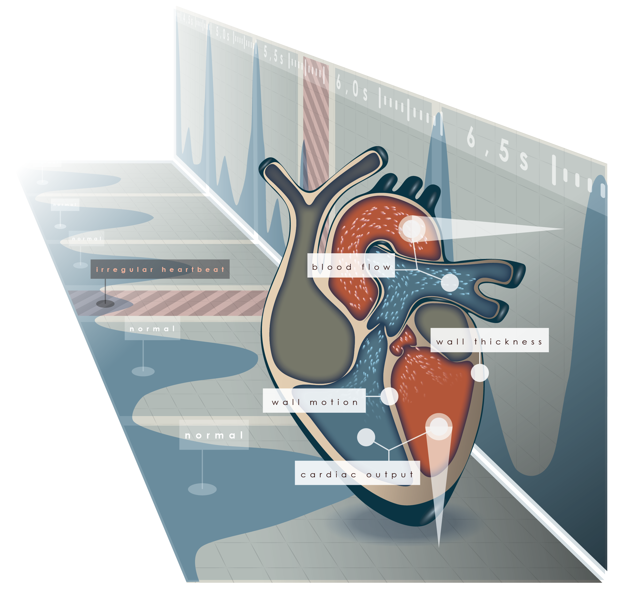
Clinical Challenges
Diagnosis of Cardiac Diseases
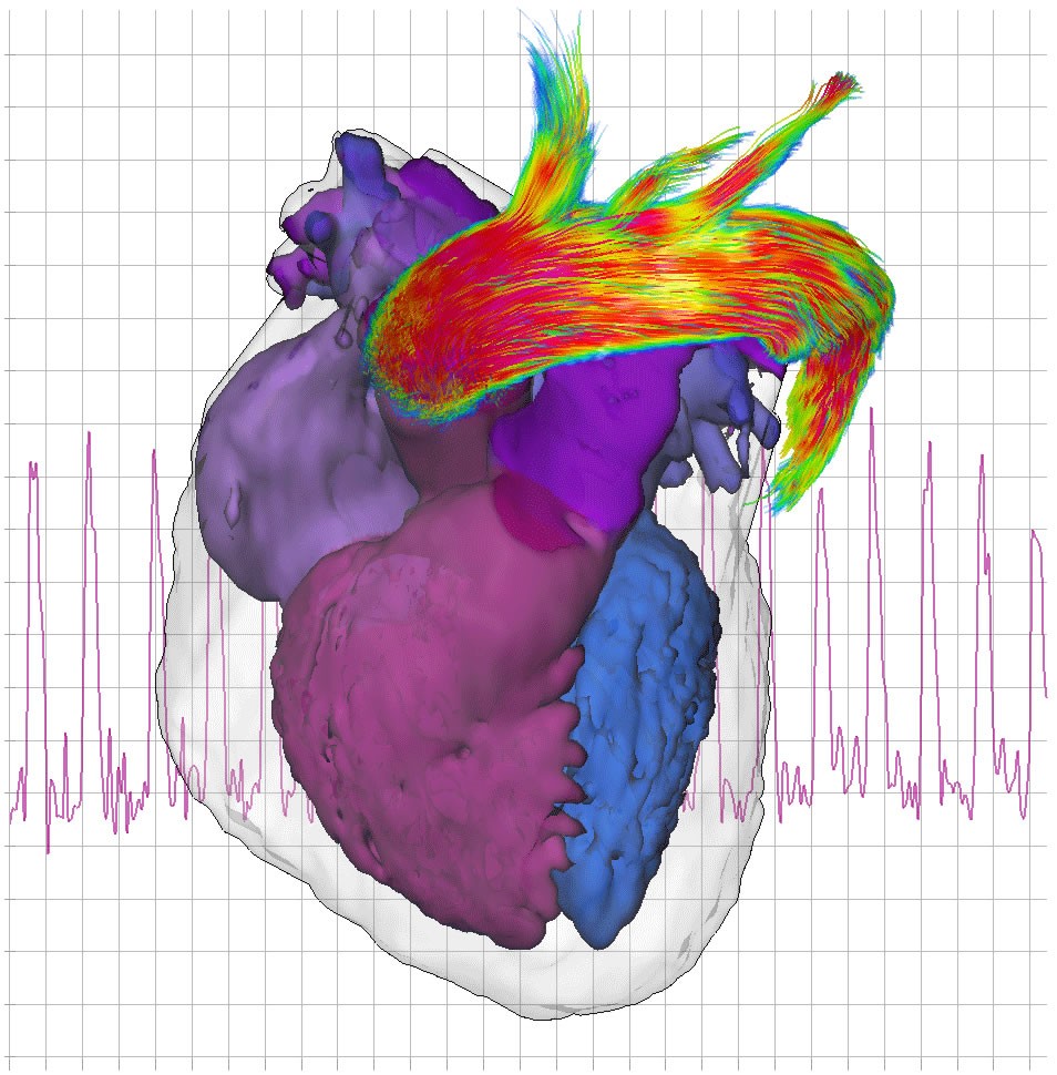
Heart failure due to congenital or acquired heart disease is affecting more than 20 Mio. people worldwide.The causes and disease pathways are however varying and have to be determined to identify the best patient-specific treatment options. Current imaging modalities such as MRI, CT and ultrasound provide the means to examine global and local cardiac bloodflow and function, perfusion, the coronary arteries as well as several aspects of the myocardial tissue state.
Software Support for Integrated Cardiac Image Analysis
Fraunhofer MEVIS develops software solutions for multimodal cardiac image analysis and quantification. Easy-to-use analysis methods are provided for different types of image data such as coronary angiographies, perfusion sequences, T1 maps, phase contrast MRI etc. Image fusion algorithms as well as advanced reporting concepts are provided to enable an integrated assessment of the complementary information derived from the different modalities.