Automated technique compares lung screening priors

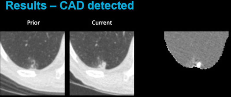
Researchers from the Diagnostic Image Analysis Group (DIAG) at the Radboud University Medical Center in Nijmegen, the Netherlands, have developed a novel computer-aided detection technique to highlight temporal changes in lung cancer screening scans. The method first performs a nonrigid registration of baseline and follow-up scan with a CT lung registration solution developed by Fraunhofer MEVIS. Subsequently, the deformed baseline is subtracted from the follow-up, thereby highlighting temporal changes such as growing lung nodules. The method has the potential to make screening follow-up easier and less tedious, emphasizes DIAG researcher Colin Jacobs, PhD, in an article on AuntMinnieEurope (http://buff.ly/1OAHV5V - Login required)
Fraunhofer MEVIS and its strategic partner DIAG have a long history of collaboration. At DIAG, the lung CT registration algorithm developed by Fraunhofer MEVIS has already been successfully applied to more than 10000 CT scans from various lung screening trials.
More information on the lung registration solution can be found here: http://www.mevis.fraunhofer.de/index.php?id=701
 Fraunhofer Institute for Digital Medicine MEVIS