 Fraunhofer Institute for Digital Medicine MEVIS
We Significantly Reduce Recall-Rates and Complication-Risks in Minimally Invasive Interventions
Targeted Navigation and Precise Motion Control
Our motion compensation as well as our catheter navigation combine a patient model with real-time sensor data to assess the therapy situation.
Titel der Webseite
Minimally invasive interventions play a central role in medicine, seen in applications including radiotherapy, heart catheters, and keyhole surgery. The Fraunhofer Institute for Digital Medicine MEVIS is researching methods to improve minimally invasive treatments significantly.
Surgery isn’t always about scalpels and open wounds. Sometimes it is possible or even necessary to employ minimally invasive methods, of which there are many varieties. During keyhole intervention, the surgical wound is made as small as possible, after which endoscopes help look inside the body and perform the operation. Some tumors can be destroyed with thin electrical needles inserted from the outside. In cardiovascular diseases, physicians navigate catheters through blood vessels, for example, to widen constricted coronary arteries. In addition, methods such as radiotherapy or focused ultrasound do not involve surgical incisions at all. For these methods, radiation or sound waves enter the body from outside and focus on a tumor to destroy it as fully as possible. Ideally, the surrounding healthy tissue remains intact.
“Minimally invasive methods take an approach of doing as little damage as possible while accomplishing the therapy goal,” says Jan Strehlow, computer scientist at Fraunhofer MEVIS. However, these gentler methods pose a challenge: During the procedure, physicians have no direct view of what is occurring in the patient. They depend on images from imaging techniques, such as X-rays or ultrasound, for both executing the procedure and monitoring the results.
Fiber optics help navigate
An example: a doctor performing a catheter intervention in the coronary vessels must X-ray their patient almost continuously. Only by using this “X-ray video,” combined with the administration of a contrast medium, can the doctor be sure that the tip of the catheter is guided correctly towards the target. This is no simple task, however, because the current three-dimensional catheter position is only shown in 2D on the X-ray image. Moreover, the method is accompanied by a considerable dose of radiation resulting from the large number of X-rays. These pose a burden not only for the patients, but also for the personnel exposed to this radiation daily.
Fraunhofer MEVIS is working on methods to reduce radiation and simplify navigation. In one of these methods, the catheter is combined with a special glass fiber. When laser light is guided into these fibers, the fiber reflects different colors of the light depending on how the fiber is bent. The spectrum of the reflected light can thus be used to calculate the curvatures along the fiber.
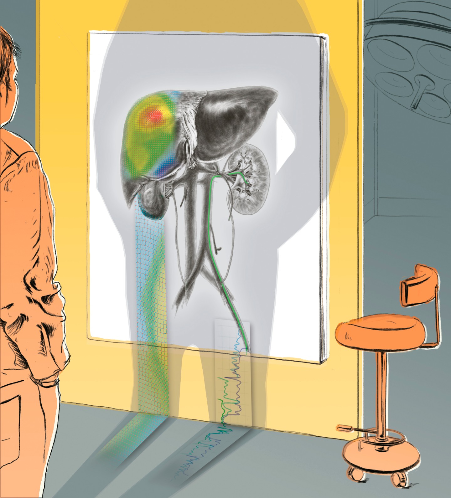
The vision: During the procedure, curvature sensor values are compared with a digital model of the patient’s vascular system obtained beforehand using MRI. “We can determine the position in the vascular system that best explains the measured catheter curvatures,” says Strehlow. This allows the physician to see the catheter moving through the vascular labyrinth on a monitor - in real time and in 3D. “We have successfully evaluated our method in a silicone model of part of a patient,” reports Strehlow. “Now we want to test it on animal models and continue development.” One conceivable application is more precise and gentler navigation in neurology, for example, to treat aneurysms.
Algorithm compensates for breathing
A further challenge is minimally invasive treatment of organs that move with the heartbeat and respiration, such as liver and lungs. “With focused ultrasound for liver cancer, you have to compensate for the movements of the patient so that the ultrasound waves always hit the tumor and not the surrounding tissue,” explains Fraunhofer researcher Michael Schwenke. In some cases, the patient lies during treatment in an MRI scanner that takes several images each second. This makes it possible to track liver movement with each breath - but only partially and with a delay.
To improve this procedure, Fraunhofer MEVIS is developing an algorithm to predict the liver’s position in upcoming moments based on MR or ultrasound images. “We are currently transferring the technology that we successfully developed for focused ultrasound to other applications such as radiotherapy or needle-based intervention,” says Schwenke. “For example, we have already created a kind of traffic light for liver biopsies that shows personnel the patient’s current breathing state.” Furthermore, the team is working on software to compensate larger patient movement, such as body rotation.
Reduce Recurrences In Thermal Ablation
")
The software assistant for interventional radiology (SAFIR) helps assessing the risk for recurrences by comparing pre- and post-interventional images through image registration and display of safety margins by a traffic light color scheme.
Reduce Recall-Rates By Personalized Device Selection In Stenting Procedures

The success of minimally invasive interventions depends on the fit and the placement of the supporting medical device like stent, valve, ...
Reduce Complexity And Radiation Exposure In Endovascular Navigation

Navigating catheters and guide-wires through the blood vessel system is often challenging and associated with high radiation doses for patient and medical staff. We develop a 3D navigation system to make endovascular interventions less complex and more safe.
A 3D navigation system can be used to visualize the position and orientation of a catheter or guide wire within patients anatomy to enable safer and more straightforward interventions. While some of our solutions target a close integration into current imaging systems, we also develop radiation-free approaches to localize the catheter during navigation.
IntelliCath
This is a demo of our novel catheter navigation system IntelliCath. A blue catheter is inserted into a simple vessel phantom to demonstrate the real-time localization displayed on the computer screen. Knowing the position of the catheter tip, we can calculate a virtual angioscopy view. The system is insensitive to motion of the whole phantom and also to deformations of the vascular system.
Minimize Undertreatment By Compensating Motion

The image stream showing organ motion is analyzed and data can then be mapped inbetween a fixed planning state and the moving motion states, including temporal predictions for real-time applications like therapy control.
Real-time images showing the motion of the object (moving state) are analyzed and data can be shown mapped to a reference state. Planning data (yellow cross) can be mapped to the moving state at all times (red cross). Predictions can be computed for real-time therapy control (orange cross).