Fraunhofer Institute for Digital Medicine MEVIS
This BMBF-funded project (funding code: 13GW0228C) began in October 2017 and will conclude in October 2020. In addition to Fraunhofer MEVIS, the consortium includes the Institute for Robotics and Cognitive Systems at the University of Lübeck, the Medical Laser Center Lübeck GmbH and the Clinics for Surgery and Radiology at the University Hospital Schleswig-Holstein in Lübeck.
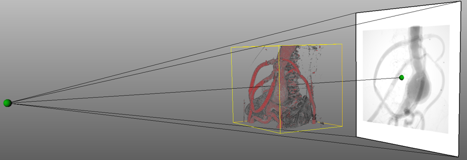
In western industrialized nations, cardiovascular diseases, such as abdominal aortic aneurysm, (AAA) are some of the most frequent causes of death. In 80% of cases, AAAs are symptom free and are usually detected only by chance. When AAAs are detected, they are treated using an endovascular aortic repair (EVAR) procedure, in which a stent graft is placed in the aneurysm region. Before the procedure, a preoperative 3D CT scan is made to analyze the position and size of the aneurysm and to prepare the surgeon. Nowadays, endovascular aneurysm repair (EVAR) procedures are conducted with 2D fluoroscopy and conventional digital subtraction angiography for catheter guidance. These image modalities have several drawbacks: They require X-ray exposure and, in addition, potentially kidney damaging contrast agent is used to visualize the vessel volume. Moreover, the depth information is missing. Thus, different technologies are being evaluated to answer the following questions:
- How can the position and shape of a catheter be determined without using X-ray imaging and contrast agent?
- How can images and data be visualized alongside the catheter position during the operation to be suitable for the surgeon?
- What accuracy can be achieved, and can we forgo X-ray imaging by adopting other technologies?