Physiological Imaging

Innovative Technologien für die Entwicklung robuster physiologischer Bildgebungsmethoden, die die klinische Diagnose unterstützen.
© Fraunhofer MEVIS
Fraunhofer MEVIS develops flexible solutions to generate robust physiological imaging parameters that support clinical diagnosis and decision making.

The vast majority of clinical decision making relies on structural images. However, physiological image information would add a new perspective, allowing earlier diagnosis and better therapy monitoring by exploiting the fact that physiological changes precede changes in tissue structure. At present, physiological imaging techniques are rarely used in clinical practice due to their lack of robustness and increased complexity. Fraunhofer MEVIS aims for reducing the complexity of these imaging techniques and making it useful for wider applications, initially focusing on Ultrasound (US) and Magnetic Resonance Imaging (MRI) techniques. We build on a long tradition in developing and deploying physiological imaging techniques by being among the leading groups in the development of Arterial Spin Labeling (ASL) sequences, a perfusion imaging method that does not require contrast agents.


Dynamic / Physiological MRI

Clinical MRI sees a general need for reliable diagnostic information from physiological imaging. However, due to the complexity of the underlying imaging techniques, problems can arise in the acquisition, the reproducibility and comparability of the output data. Reducing the variability in physiological imaging data will make the outcomes more robust and comparable and ultimately lead to earlier detection of diseases, as well as robust quantification and therapy monitoring in the field of neurodegenerative diseases. We build on existing, in-house technology for adaptive, real-time MRI scanning to modularly generate one-click solutions in our scanner agnostic MRI framework gammaSTAR that reduce complexity and increase harmonization of imaging data in this promising field.


Motion Correction

Physiological imaging techniques often use subtle differences between multiple timepoints to detect changes in physiological characteristics. Subject motion can easily lead to misregistered images which will result in loss of information. During the last years we developed retrospective and prospective motion correction techniques that reduce the effect of subject motion on the image outcome, e.g. in arterial spin labeling, diffusion MRI and functional MRI.

Automated quality assessment of MR image series.
© Fraunhofer MEVIS
Automatic image quality assessment can rate imaging data related to general image quality or specific artefacts. This can be utilized retrospectively in large imaging studies, but also for online-control at the MRI scanner.

Online Image Quality Observation

Low signal-to-noise and a general lack of image quality are common challenges of physiological MRI sequences. Our automated image quality assessment methods can help with observing the image quality for retrospective decision making or for applying dynamic changes to the MRI sequence during the scan to maintain a high image quality.

© Fraunhofer MEVIS
Our time-encoded pseudo-continuous arterial spin labeling (ASL) sequences in combination with single-shot 3D-GRASE readout provide shorter scan time and higher SNR than conventional ASL methods.

Patient-Specific Adaptive Imaging

Each individual has a different physiology. These often subtle differences have an influence on the MRI measurement. Fraunhofer MEVIS therefore develops adaptive, patient-specific imaging algorithms. For each patient, the parameters of the physiological MRI sequence should be automated and individually adapted. Time-consuming preparation of the imaging parameters prior to the scan would then be avoided.

Automated Post-Processing 

Valuable information from physiological images can typically only be obtained during post-processing. At present this is often done on external computers and is time-consuming. We aim to connect post processing directly to the measurement, so that valuable waiting time for evaluation results is eliminated.

© Fraunhofer MEVIS
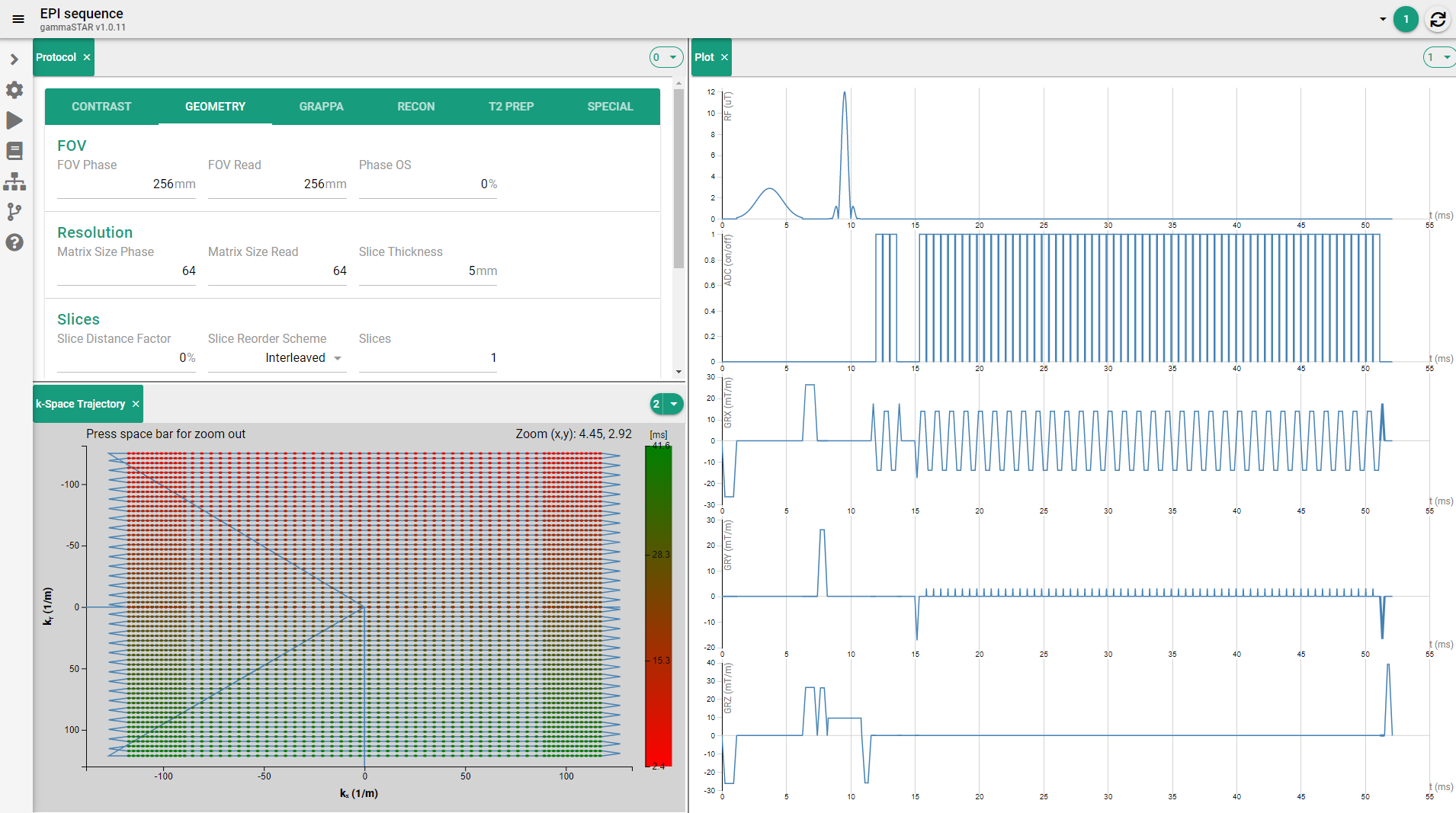
gammaSTAR offers a user-friendly web-frontend for pulse-sequence development with on-the-fly plotting and other convenient features as for example a k-space trajectory view.

Scanner Agnostic MRI Sequences

Our scanner-agnostic MRI framework gammaSTAR provides a generalized format for MRI sequence generation, enabling cross-vendor and cross-scanner portability of MRI techniques to overcome the lack of cross-scanner integration in today’s marketplace. It allows more efficient sequence development and implementation, providing a breakthrough in MRI image harmonization. Applied to physiological imaging techniques, the framework can help reduce the variability of image data across different devices. It is developed in a way that allows easy and modular exchange of imaging modules, accelerating the integration of the above techniques into the clinical workflow.


Get more information about gammaSTAR

Visit our open web frontend and explore gammaSTAR:  https://gamma-star.mevis.fraunhofer.de


Research Highlights (Selection)

  • Cordes C, Konstandin S, Porter D, Günther M. Portable and platform‐independent MR pulse sequence programs. Magn Reson Med (2020) 83:1277–1290
  • Hoinkiss DC, Erhard P, Breutigam N-J, von Samson-Himmelstjerna F, Günther M, Porter DA. Prospective Motion Correction in Functional MRI Using Simultaneous Multislice Imaging and Multislice-to-Volume Image Registration. NeuroImage (2019) 200:159-173
    (DOI: 10.1016/j.neuroimage.2019.06.042
  • Hoinkiss DC, Porter DA. Prospective Motion Correction in Diffusion-Weighted Imaging Using Intermediate Pseudo-Trace-Weighted Images. NeuroImage (2017) 149:1-14.
  • von Samson-Himmelstjerna F, Madai VI, Sobesky J, Guenther M (2016) Walsh-ordered Hadamard time-encoded pseudocontinuous ASL (WH-pCASL). Magn Reson Med 76(6):1814–1824

Conference Contributions (Selection)

  • Breutigam NJ, Hoinkiss DC, Buck MA, Mahroo A, von Samson-Himmelstjerna F, Guenther M (2020) Optimal subject-specific pCASL settings by automated inner-scan timing adaption. Proceedings of the 28th Annual Meeting of the ISMRM. 3282
  • Huber J, Hoinkiss DC, Guenther M (2020) Subject-specific background suppression in 3D pseudo-continuous arterial spin labeling perfusion imaging. Proceedings of the 28th Annual Meeting of the ISMRM. 3283
  • Hoinkiss DC, Struckmann S, Werner N, Schmidt CO, Günther M, Hirsch J (2020) Long-Time Stability of Image Quality in the German National Cohort (NAKO) Imaging Study Based on Automatic Image Quality Assessment. Proceedings of the 37th Annual Scientific Meeting of the ESMRMB. L01.124
  • Hoinkiss DC, Cordes C, Konstandin S, Huber J, Wilke RN, Günther M. (2019) Real-Time Sequence Control for Prospective Motion Correction in a Dynamic, Platform-Independent MRI Framework. Proc ESMRMB. S14.02
  • Günther M (2018) Arterial Spin Labeled Input Function (ASLIF): signal acquisition during pseudo-continuous arterial spin labeling. Proceedings of the Joint Annual Meeting of the ISMRM and the ESMRMB. 0305

Dynamic / Physiological Ultrasound Imaging

Deep Learning based ultrasound image reconstrucion may enable high quality image data from less input data.
© Fraunhofer MEVIS
Fraunhofer MEVIS develops deep learning (DL) methods that allow reconstructing ultrasound high quality images from less input data, which speeds up acquisition times when monitoring processes in the body. Here, a deep learning network was trained to produce higher quality (multi-shot) images from the data of a single planewave shot.

In thermal therapies a temperature change is induced into (malignant) tissue in order to destroy it or to promote healing or can be used in drug delivery. The therapeutic tools in this domain (e.g. HIFU, RF-, cryo-, laser-ablation) allow for a localized, targeted delivery, but they are applied without direct visual contact to the target regions. The therapy devices themselves usually do not bring the visualization tools for treatment monitoring, when however in these therapies dynamic monitoring is absolutely crucial. Monitoring is used to ensure the correct dose is delivered to the right place, sparing surrounding healthy tissue. In particular it is important to see the dynamic change (e.g. in temperature) before the structural change (e.g. ablation) occurs. Often MRI is used as an expensive monitoring device for localized temperature changes. Not only does a large MRI system dominate the setup around the patient, but it may also not always achieve the necessary spatial resolution and framerate for some procedures. Ultrasound imaging could serve as an inexpensive and mobile tool to assist in treatment monitoring in real-time. However, here ultrasound itself is still facing practical problems in its robust applicability.


Fraunhofer MEVIS is working on bringing ultrasound imaging to robust application in therapy monitoring:

  • Thermal Therapy Monitoring
    Fraunhofer MEVIS investigates the use of ultrasound based methods for dynamic imaging in thermal therapies, in particular the use of deep learning in approaches for US-monitoring thermal therapies (with application in HIFU).
  • Accelerated Image Reconstruction
    Fraunhofer MEVIS develops algorithms to increase the acquisition speed for ultrasound imaging by allowing the reconstruction of images from less acquired data. Gains in image acquisition speed makes dynamic imaging more robust to motion artefacts which occur as a result of regular and irregular patient motion. It can also be used to enable more data consuming acquisition schemes (e.g. 3D imaging) to achieve real-time performance.


Research Highlights

Literature

  • Rothlübbers S, Strohm H, Eickel K, Jenne J, Kuhlen V, Sinden D, Günther M (2020) Improving Image Quality of Single Plane Wave Ultrasound via Deep Learning Based Channel Compounding Proc IUS 2020
  • Strohm H, Rothlübbers S, Eickel K, Günther M (2020) Deep learning-based reconstruction of ultrasound images from raw channel data. Int J CARS

Conference Contributions

  • Strohm H, Rothlübbers S, Eickel K, Günther M (2020) Beyond Classical Ultrasound Contrast via Deep Neural Network at IUS 2020
  • Fournelle M, Hewener H, Speicher D, Rothlübbers S, Schwenke M, Jenne J, Bücker A, Tretbar S (2020) MR-compatible ultrasound for improved biopsy needle guidance at BMT 2020
  • Strohm H, Rothlübbers S, Eickel K, Jenne J, Kuhlen V, Sinden D, Günther M (2020) Applications of Deep Learning in Ultrasound Image Reconstruction at BMT 2020