 Fraunhofer Institute for Digital Medicine MEVIS
Risk Analysis and Resection Planning
Solutions & Technologies
For more than 15 years, Fraunhofer MEVIS has supported surgeons in planning of complicated liver surgeries [1]. Resection proposals and risk analyses that consider patient-specific liver anatomy have changed surgery and improved the safety and outcomes of such interventions [2,3].
There are several risks during liver surgery, with liver failure being the most severe. Major risk factors include the volume of the remnant liver after surgery as well as the health of the remaining liver tissue. For oncologic resections, the tumor safety margin presents another risk and is a critical marker for cancer recurrence and the success of the surgery. These risk factors, added to the complexity of the liver vasculature, motivated the development of a patient-specific, computer-assisted planning platform at Fraunhofer MEVIS. The software analyses radiological images and calculates information on the drainage and perfusion of the organ using mathematical models. The algorithms quantify risks for the intervention and generate a detailed 3D visualization of the liver and its vascular systems. Supply areas of these blood vessels, such as the portal vein and hepatic arteries, are calculated and help to evaluate and optimze the surgical planning.
Privacy warning
With the click on the play button an external video from www.youtube.com is loaded and started. Your data is possible transferred and stored to third party. Do not start the video if you disagree. Find more about the youtube privacy statement under the following link: https://policies.google.com/privacyData Analysis
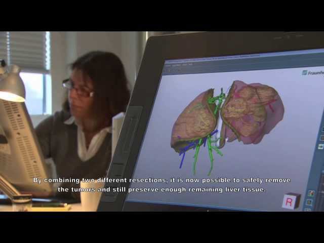
Based on contrast-enhanced CT or MR images, the organ, vessels structures, and lesions are aligned from the different scans and reconstructed in three dimensions. Supply and drainage territories are computed from the individual vascular systems of portal and hepatic veins.
Risk Analysis
Vessels in different safety margins of the tumor and their depending territories are determined to estimate the surgical risks and optimize resection proposals. User- defined virtual resections can be combined with supply and drainage territories to evaluate the remaining functional volume of the remnant liver [1,4].
Visualization of Results
Anatomical and pathological structures, computed liver territories, color-coded risk branches, and areas with potentially obstructed outflow can be visualized in many ways, rotated in 3D, and explored interactively. Volumes of organ, lesions, remnant liver, and risk territories are displayed on demand to support choosing the best surgical strategy [5].
Transfer into the OR
Predefined collections of results can be exported for use in a dedicated navigation system or displayed on a mobile device in the operating room. Additionally, the Fraunhofer MEVIS Liver Explorer app offers tools to update resection volumes during surgery, measure distances and vessel structures, and erase vascular branches on a fingertip.
Highlights
- Analysis of CT and MRI data
- Assessment of supply and drainage territories
- Dedicated risk analysis for tumor surgery and LDLT
- More than 7000 cases analyzed
- Liver Viewer and 3d-pdf for exploration of results
- Dedicated export for use in a navigation system
- Mobile Liver Explorer App for use in the OR
- Software that modifies surgical strategy
[1] Schenk A et al. IEEE Pulse 2011. [2] Lang H et al. Arch Surg 2005 [3] Wang Y et al. Dig Surgery 2012. [4] Hansen C et al. IJCARS 2014. [5] Endo I et al. In Hilar Cholangiocarcinoma 2013.