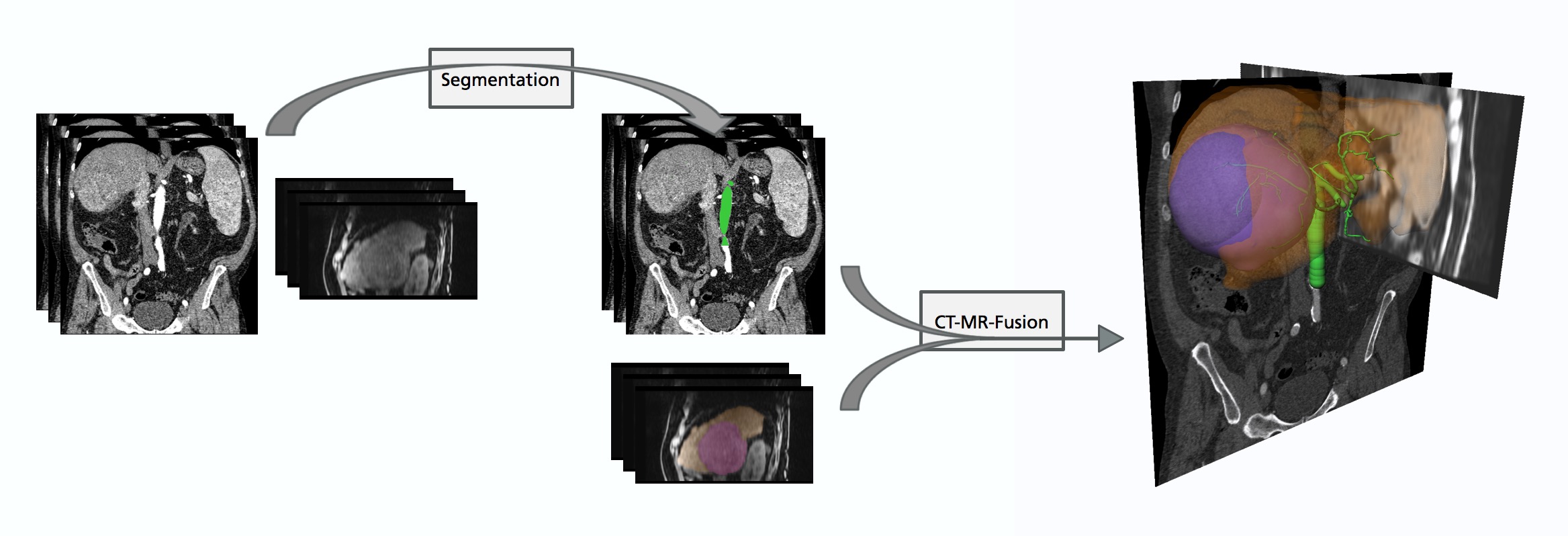
A variety of medical images can be involved in diagnosis, treatment planning, treatment assessment, and follow-up assessment. In order to enable the combined use of information from different image modalities and capture times or to assess changes, image registration is a key technology in image-based medicine. In particular, abdominal images in the field of liver surgery provide a wide spectrum of image registration applications.
Registration of Abdominal Images
Liver Motion Correction and Abdominal Follow-up Image Registration
In medical images, the position of the liver is strongly influenced by the patient breathing. Therefore, a comparison or fusion of abdominal images is often hampered by different organ positions. Deformable motion correction and deformable image registration provides a solution to this issue. The goal is to harmonize the images obtaining physically reasonable results.
Anatomical correspondence of multi-modal abdominal images is achieved by deformable image registration. This enables local position correlation.
Registered abdominal follow-up images enable position synchronization through the whole image volumes.
Multi-Modal Abdominal Image Registration
Image fusion of multi-modal abdominal images enables the use of combined information. In the case of hepato-cellular cancer, contrast-enhanced MR images allow to differentiate between normal liver parenchyma and liver lesions. This enables liver volume and tumor burden calculations. Based on deformable image registration of T2-weighted MRI, a precise lesion characterization becomes easier and more convenient. For cancer treatment the patient's liver performance is a decisive factor. The local liver function can be provided by registered diffusion-weighted MRI. The vessel system of the liver can be derived from CT images and enables the planning of surgical resections, catheter-based interventions, and the consideration of cooling effects for radio-frequency ablation. Radiation-based therapies also use the Hounsfield values from the CT images in order to predict the therapeutic effect.
Anatomical correspondence of multi-modal abdominal images is achieved by deformable image registration. This enables local position correlation.
Intra-Operative Navigation and Real-Time Tracking
Visualization of the registered 2D ultrasound slice and the 3D CT image volume highlighted by the arterial and venous vessel systems.
Pre-operative liver surgery planning is typically performed some days prior to the intervention based on abdominal CT of the patient. This involves the segmentation of vessels, liver segments, the definition of intersection lines, and a risk calculation. During the intervention the surgeon is guided by tracked ultrasound images of the liver. To fully exploit the pre-operative planning, the CT data has to be fused to the actual deformation of the liver given by the ultrasound data.
 Fraunhofer Institute for Digital Medicine MEVIS